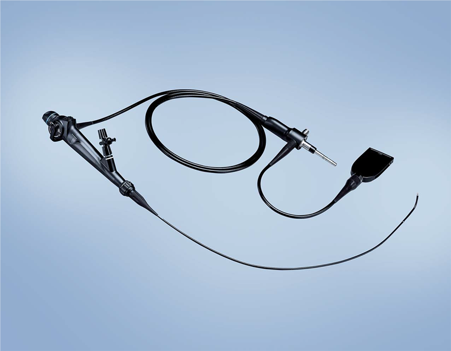
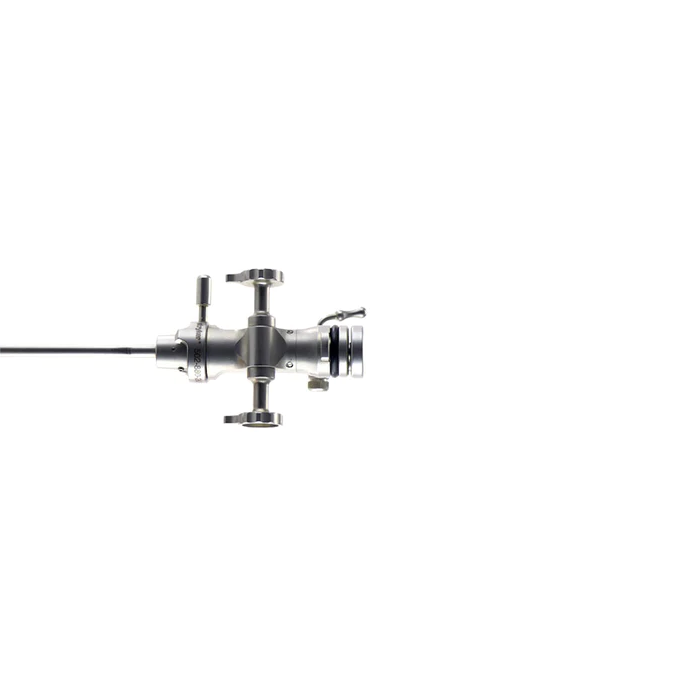
Olympus URF-V Flexible Video Ureteroscope
See what you have never seen before.
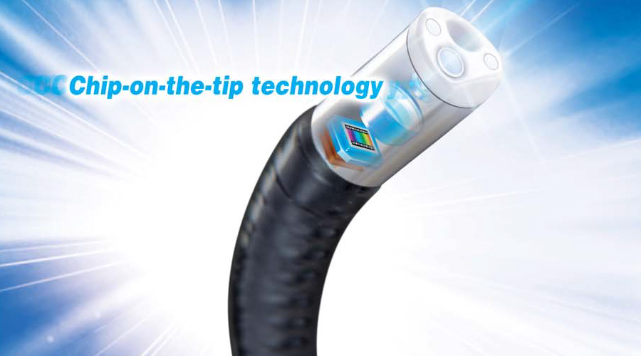
"EYE inside" technology
The CCD imaging sensor in the distal end provides a moiré-free, bright image with high color reproduction without the need for a camera head attachment. The use of the integrated distally located CCD imaging sensor eliminates the need to focus.
Largest image size
Olympus ureteroscopes boast the largest image size** currently available. The URF-V's image is about three times larger than that of our conventional fiberscope, making the most of its high-resolution image quality and enhancing observation.
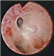
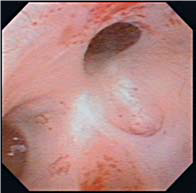
Conventional fiberscope image* The image of URF-V
* The OES uretero-reno fiberscope URF-P5 connected with the camera head OTV-S7H-1D-L08E.
**As of November, 2008.
Narrow Band Imaging
NBI helps in the observation of mucosal morphology; NBI works by altering the white light source to consist of specific wavelength bands, which take advantage of the scattering and absorption properties of human tissue. This provides improved visual contrast of the surface structure and fine capillary patterns of the mucous membranes, which are normally difficult to distinguish. NBI takes advantages of the characteristics of the light that penetrates the mucosa by depicting capillaries in the superficial layer of the mucosa more clearly than with conventional white light.
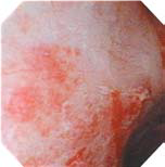
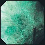
Conventional image NBI image
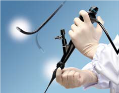
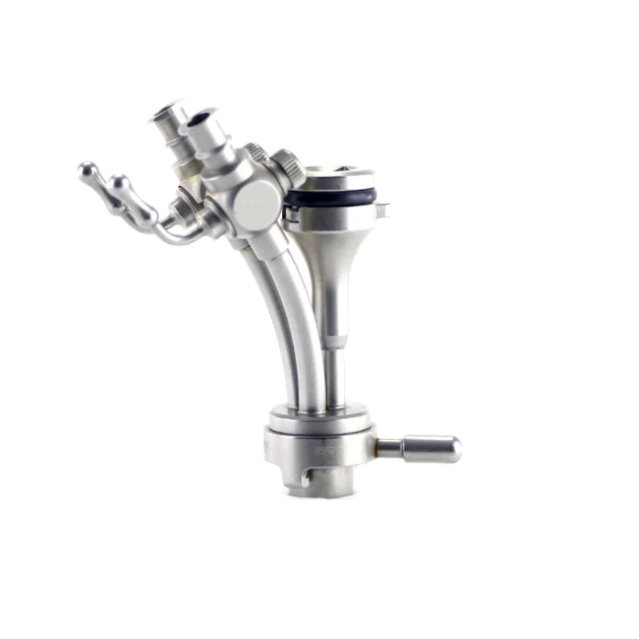
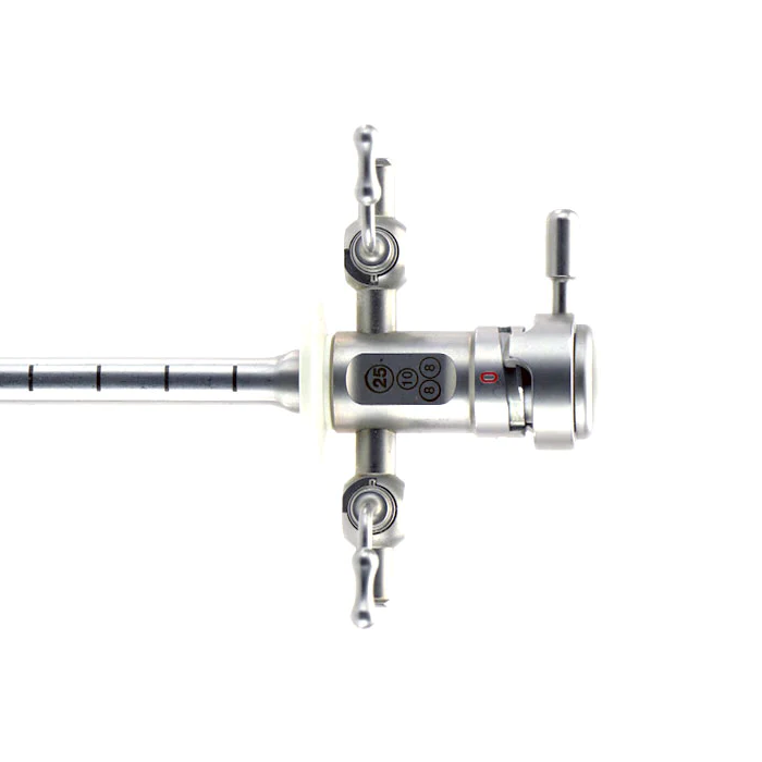
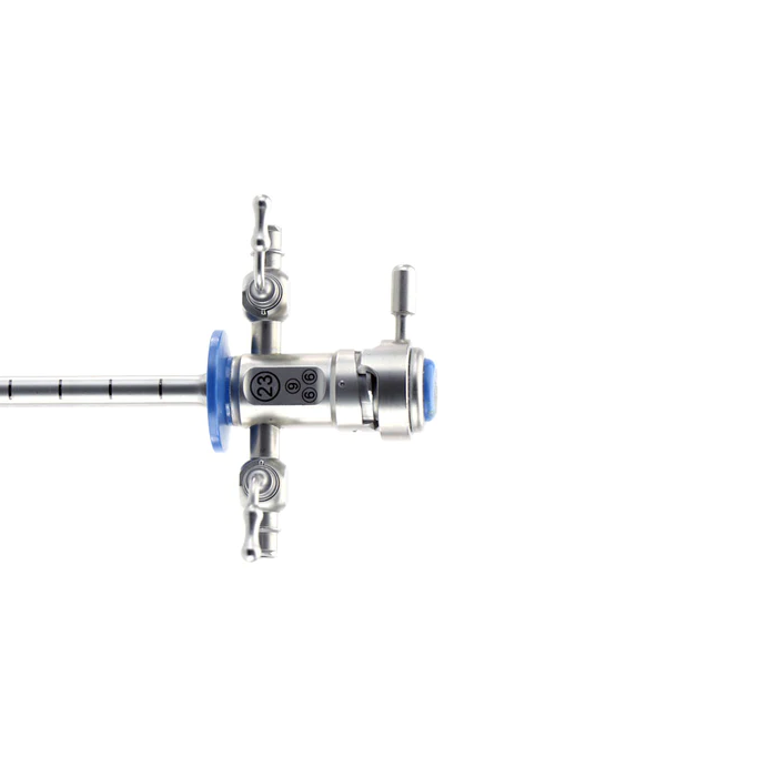
Smooth Handling
A new insertion tube rotation function with a rotation angle of ±90 degrees enables fine adjustment of the laser tip position when aiming at the stone,while also allowing a relaxed working position
UP 180° /DOWN 275°
The 275° down angulation enables optimal visualization in the lower calyx, while the 180° up angulation (with a small radius) is ideal for accessing the upper/middle calyces.
Ergonomic Design
The integrated camera and light guide cable assures easy scope handling during the procedure. Four programmable buttons on the control section are always within your reach, providing quick access to image capture, white balance, zoom and other frequently used functions.


 Microsystem
 Endoscopysystem
 Energysystem
 EndoscopyConsumables
 +86-21-54286005
 Room 602, Building 1, No. 111 Luxiang Road (Greenland Park Plaza), Baoshan District, Shanghai, China
  English
 中文
中文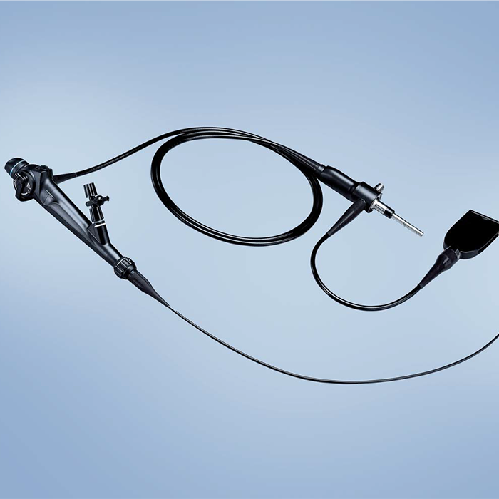
 URF-V_Brochure_EN


 中文
中文 English