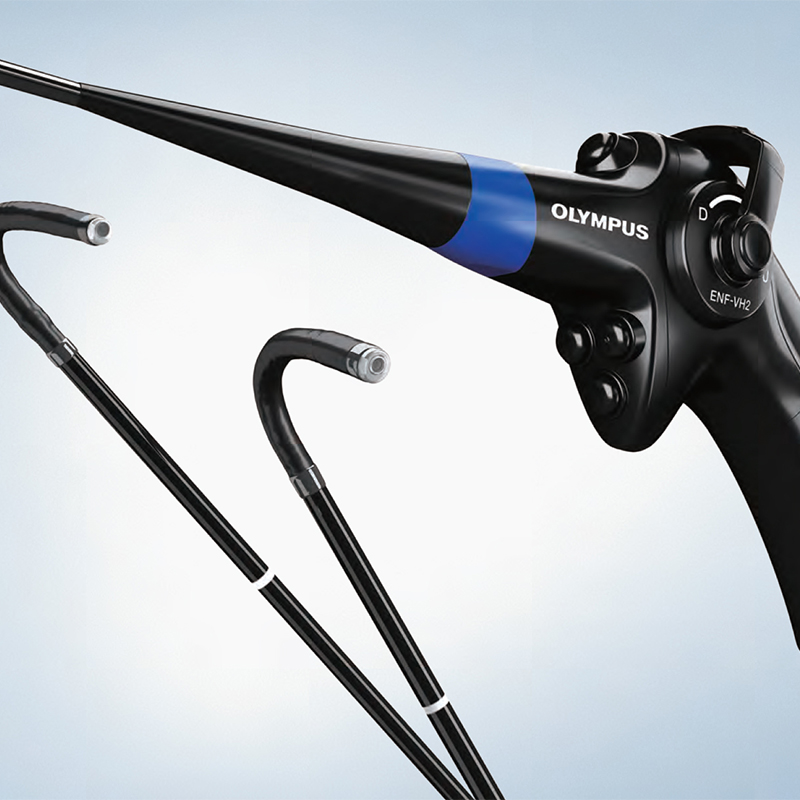
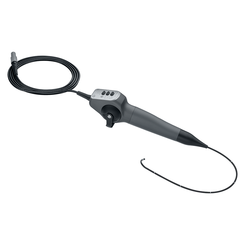
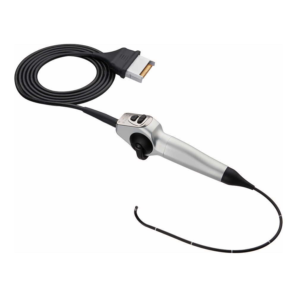
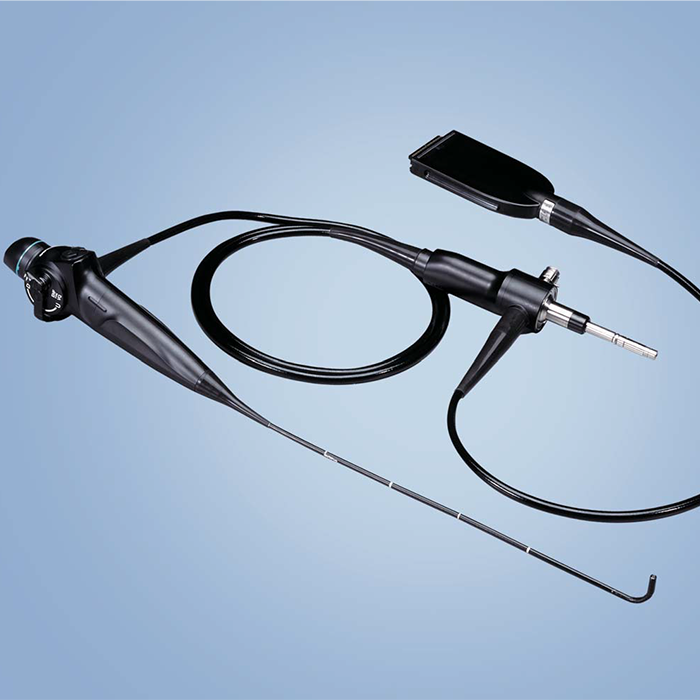
Olympus ENF-VT3 Flexible Video Rhinolaryngoscope
4-Angle Observation and Treatment with High-Quality Imaging

Four-Direction Angulation
Featuring the same slim outer diameter as the conventional model*, the ENF-VT3 is the world’s fi rst rhino-laryngo videoscope to incorporate four-direction angulation capability.The addition of 70° right/left angulation to the previously available* 130° up/down has dramatically improved the approach to a lesion. Angulation in four-directions is possible using just one hand, allowing the other hand to simultaneously manipulate a therapeutic accessory.


Higher-Quality Imaging
The incorporation of a highperformance CCD further improves image quality and contributes to observations and treatments of small lesions due to its clear fi eld of view.

Close Focus Observation
The close-focus capability enables observation from just 2 mm for situations when more precision and fi ner detail is required, such as viewing minute variations and lesions in the mucosa.
Enhanced Visualization
Narrow Band Imaging (NBI) is a patented optical image technology that enhances the visibility of vessels and other tissue on the mucosal surface. NBI works by fi ltering the white light into specifi c light wavelengths, which are absorbed by hemoglobin and penetrate only the surface of human tissue. This highlights areas of increased vascularity which are normally diffi cult to distinguish.


 Microsystem
 Endoscopysystem
 Energysystem
 EndoscopyConsumables
 +86-21-54286005
 Room 602, Building 1, No. 111 Luxiang Road (Greenland Park Plaza), Baoshan District, Shanghai, China
  English
 中文
中文
 ENF-VT3_Brochure_EN


 中文
中文 English