Automated Imaging for Accurate and Efficient Experiments
Designed for automated multidimensional observation and easy experiment setup, the IXplore Pro microscope system facilitates effortless data collection and seamless acquisition of panoramic, multichannel images.

Ease of Use
The Graphical Experimental Manager (GEM) of cellSens Dimension software offers fully automated multidimensional observation (X, Y, Z, T, wavelength, and positions) and eases experiment setup. To increase efficiency, you can define macro functions, such as executing deconvolution processing, in the GEM.

Precise, Reliable Hardware
Automated Focusing
The cellSens software’s multipoint focus map enables automated focusing across wide image areas with multiple objective lenses, making it easier to stay in focus while you navigate your samples.

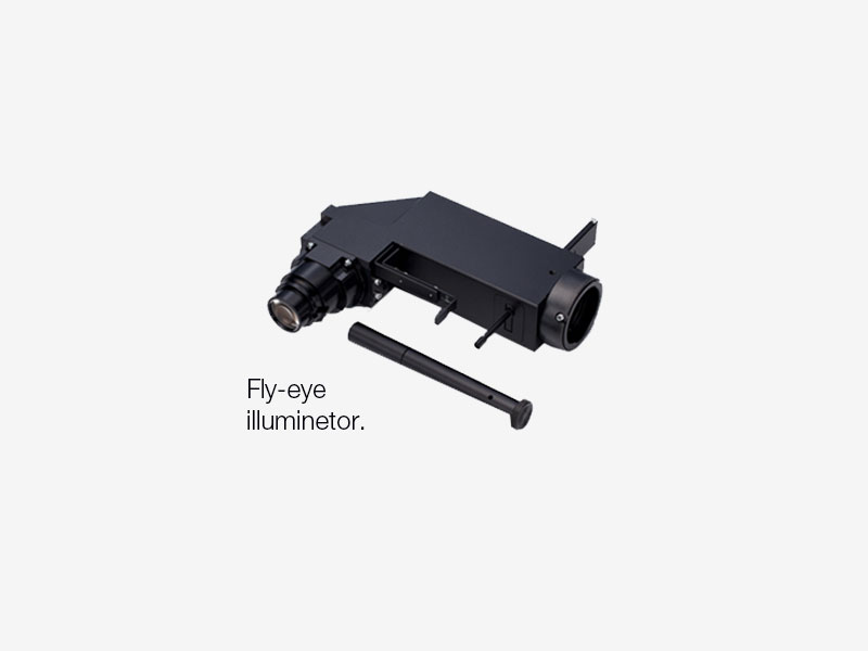
Bright, Uniform Fluorescence Illumination
The fluorescence illuminator (IX3-RFALFE) incorporates a fly-eye lens system to distribute light evenly. The system provides bright and uniform illumination to the entire field of view, which is well-suited for image stitching applications.
Well Plates Made Simple
The Well Plate Navigator and Database solutions for cellSens software facilitate proper screening during an experiment. Together, they improve the efficiency of viewing and analyzing well plate images with a large amount of data.
Information such as date, file name, or well plate number are easily selectable with icons, displaying any selection of captured images to be used for further analysis. These solutions also enable continuous analysis of selected images (the batch macro function) using the well plate GUI.


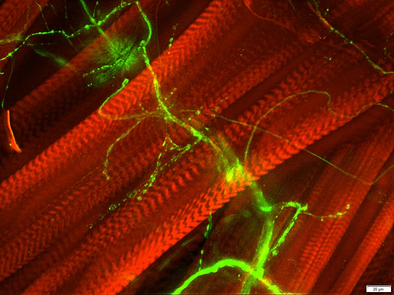
Rapid Deconvolution
Olympus cellSens Dimension software includes live 2D deblurring for preview and acquisition, enabling better focusing of thick specimens. For further detail enhancement, TruSight deconvolution is available to reassign out-of-focus light. TruSight uses a constrained iterative deconvolution algorithm to produce improved resolution, contrast, and dynamic range with industry-leading high speed through GPU processing. To improve experiment efficiency, deconvolution processing can be defined as a macro function in the GEM.

Left: Without TruSight / Right: With TruSight Left: Without TruSight / Right: With TruSight
Simplify Your Workflow
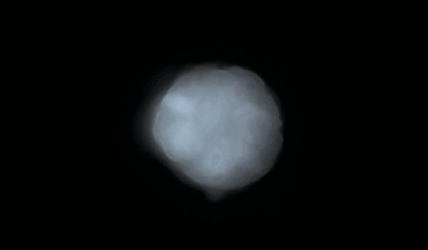
Suitable Objectives for Observation with Plastic Vessels
LUCPLFLN series objectives and, in particular, the UCPLFLN20XPH (NA 0.7) objective are well-suited for observation using plastic dishes. The objectives enable high-resolution observation of the cell proliferation process and deliver improved contrast across a wide area. This gives you the flexibility to image through plastic-bottom dishes in addition to glass.
*Image: iPS-cell expressing Nanog reporter (GFP) Image data courtesy of: Tomonobu Watanabe, Ph.D. Laboratory for Comprehensive Bioimaging, RIKEN Quantitative Biology Center


Advanced Analysis
Images can be easily converted to statistically relevant data with cellSens software. The software features region-of-interest, phase analysis, and cell counting capabilities. Export raw measurement data to Microsoft® Excel® software or a cellSens workbook with a single click.


 Microsystem
 Endoscopysystem
 Energysystem
 EndoscopyConsumables
 +86-21-54286005
 Room 602, Building 1, No. 111 Luxiang Road (Greenland Park Plaza), Baoshan District, Shanghai, China
  English
 中文
中文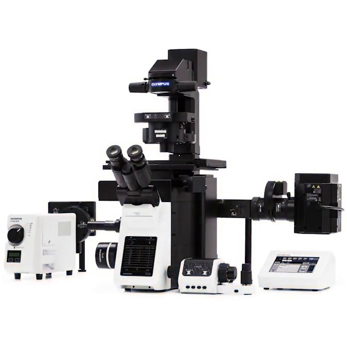
 IXplore_Brochure_EN


 中文
中文 English