 EndoscopyConsumables
 English
 中文
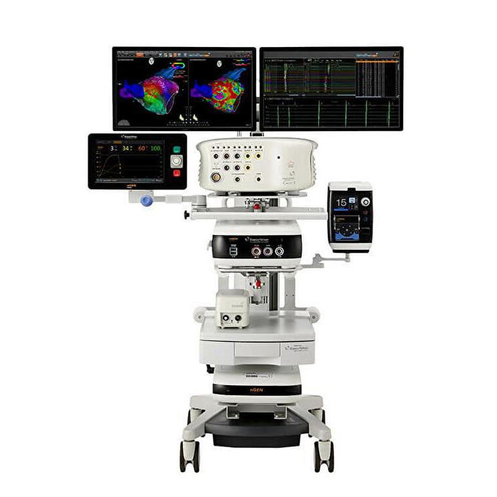
中文Biosense Webster CARTO™ 3 Cardiac mapping System
CARTO™ 3 System Version 8 advances the CARTO™ mapping capabilities by bringing advanced tools for physicians across a range of procedures.
Phone:+86-21-54286005
Resources
 CARTO™ 3_Brochure_EN


 Microsystem
 Endoscopysystem
 Energysystem
 +86-21-54286005
 info@tenmed.net
 Room 602, Building 1, No. 111 Luxiang Road (Greenland Park Plaza), Baoshan District, Shanghai, China
CARTO™ 3 System Version 8 advances the CARTO™ mapping capabilities by bringing advanced tools for physicians across a range of procedures.
Phone:+86-21-54286005
CARTO™ 3_Brochure_EN
3D NAVIGATION & MAPPING TECHNOLOGY
CARTO™ 3 System Version 8 advances the CARTO™ mapping capabilities by bringing advanced tools for physicians across a range of procedures. Through enhanced signal analysis, improved substrate characterization and utilization of ultrasound technology, version 8 provides the tools to support more efficient EP procedures, reduce procedure time and drive reproducible results.
*When using MULTIPOLAR
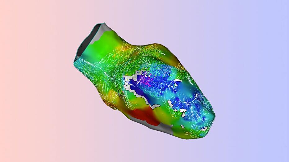
LAT Velocity Vectors (LVV) provides a representation of the activation wave velocity over a substrate map done in sinus rhythm or during pacing
Local Velocity Vectors aims to improve the specificity of localizing the most arrhythmogenic regions within the scar and reduce the need to characterize the substrate properties under different activation wavefronts.*
*Study includes sixteen patients with ischemic VT, with substrate maps obtained in SR (6), RV pacing (12), and LV Pacing (9). In the 7 patients VT was non-inducible at the end of the procedure. LAVAs were successfully eliminated in 15 of the 16 cases.
The Stability+ algorithm continuously analyzes the overall movement of the ablation catheter, separating the movement due to respiration from the actual catheter movement.*
This continuous analysis reduces the time for initial indication of stability and the appearance of VISITAG™ Module Sites.
*Compared to previous version of CARTO™ 3 System software.
The CARTO™ 3 System is based on the following three foundations:
The ECG subsystem supports receiving Body Surface ECG signals and Intracardiac (IC) Electrogram channels. It samples, filters, presents, and records ECG signals. In addition, it passes the ECG signals through to the recording system
CARTO™ Magnetic Location Technology, supplying locations based on electromagnetic fields generated by the location pad and measured by Biosense Webster catheters that have embedded magnetic sensors.
Advanced Catheter Location (ACL), supplying location data from each electrode connected to the system. The location is calculated based on the signals received from six patches attached to the patient. The variation in tissue conductivity is calibrated using magnetic location technology that is not influenced by human conductivity.
![]()
The mapping technology is used to build maps of the heart chambers, for display in the Main Map Viewer. These maps are built by combining the accurate location data and ECG data. The system enables the following mapping methods:
The CARTO™ 3 System provides several modules and optional features for differing clinical needs.
![]()
Reproducibility You Can Trust*:
*When mapping with CARTO™ 3 System Version 8.
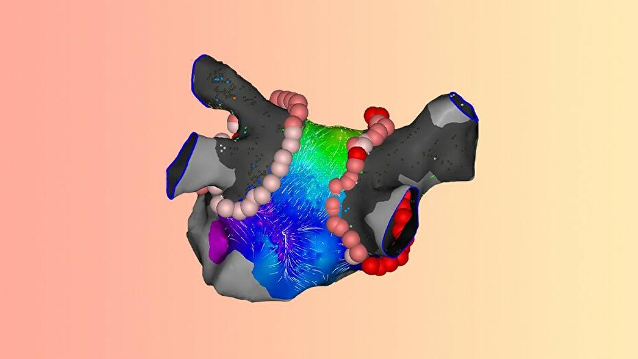
**This is a single‐center retrospective study. A total of 33 AAFLs in 27 patients were analyzed. The new tool identified an area of fractionation at the critical isthmus in 91% of the cases.
![]()
CARTOSOUND™ FAM Module reconstructs the left atrial (LA) anatomy on the CARTO® 3 Mapping System with a few rotations of your ultrasound catheter, eliminating the need for manual contouring.
*CARTOSOUND™ FAM Module uses Deep Learning, which is a subset of AI.

 中文
中文 English