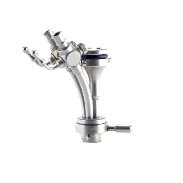
Olympus URF-V3 Flexible Video Ureterorenoscope
High Image Quality and Durability in Superslim 8.4 Fr.

Olympus’s unique DuraBend insertion-tube design reduces stress on the bending section during insertion in the narrow renal pelvis by passive bending, which increases the durability of the URF-V3.
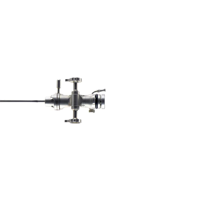
Unique Insertion-Tube Rotation for More Comfort
The 120° rotation function allows the operator to change the shaft’s angle by rotating a ring on the handle. This potentially allows for longer laser activation that makes treatment of large stone burdens feasible, more precise, and more comfortable.
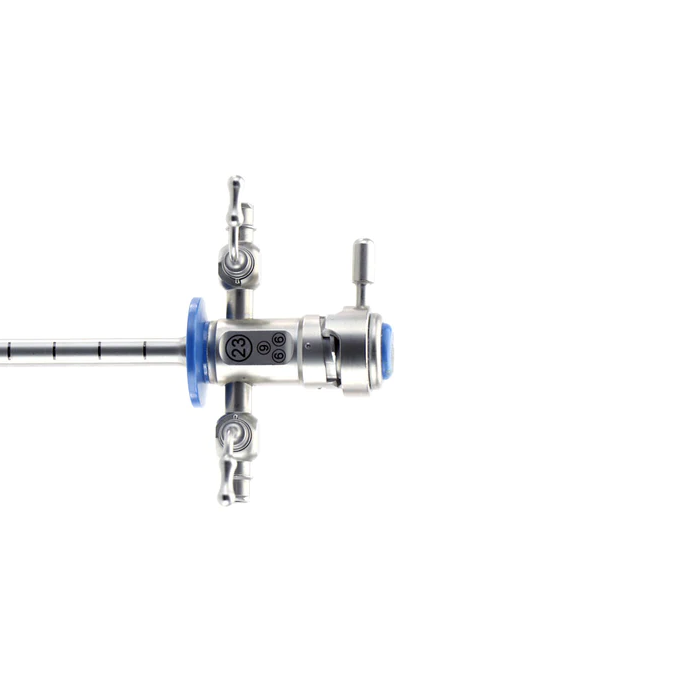
8.4 Fr. Slim Diameter Shaft
- Slim URF-V3 fits into 10/12 Fr. access sheath allowing for access in tight ureters
- The stiffness gradient of the scope allows for great torque of the scope’s shaft and a flexible tip for increased maneuverability during ureteroscopies
- Using the URF-V3 in combination with a 12/14 Fr. access sheath ensures the clearest visualization thanks to improved flow

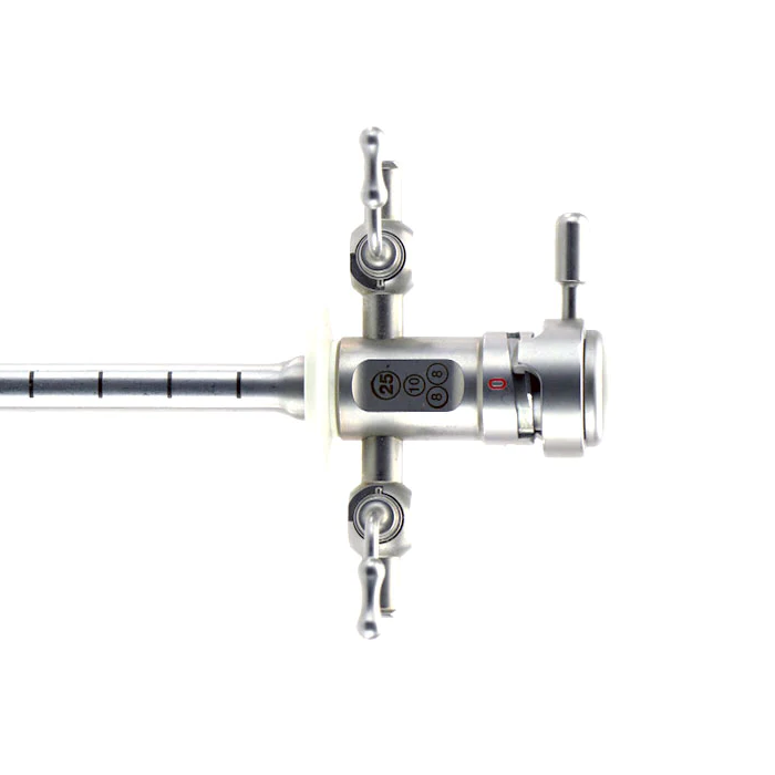
Conventional scope
(approx. 10 Fr. shaft)
275° Wide Angulation Range
Deflection capabilities of 275° up and down allow for optimal visualization of lower renal calyxes as well as upper and middle calyxes.

Stone Treatment and Tumor Detection with NBI
Stone Treatment
The URF-V3 provides a large, distortion-free digital image and an even illumination of the high-resolution image. The ability to resolve small stone fragments in a well-illuminated image ensures that the surgeon can fulfill requirements of thorough and efficient treatment.

Fiberscope URF-P7 Videoscope URF-V3
Tumor Detection
Narrow Band Imaging (NBI) as optical image enhancement technology provides substantial help in better identifying upper tract tumors due to improved visibility of vessel structures.

White light NBI


 Microsystem
 Endoscopysystem
 Energysystem
 EndoscopyConsumables
 +86-21-54286005
 Room 602, Building 1, No. 111 Luxiang Road (Greenland Park Plaza), Baoshan District, Shanghai, China
  English
 中文
中文
 URF-V3_Brochure_EN


 中文
中文 English