| Power Supply
|
Rated voltage
|
100-240 V AC; Within ±10%
|
|
Frequency
|
50/60 Hz; within ±3 Hz
|
|
Rated input
|
600 VA
|
|
Size
|
Dimensions (W x H x D)
|
370 x 198 x 488 mm; 398 x 218 x 580 mm (maximum)
|
|
Weight
|
19.4 kg
|
|
Classification (Medical Electrical Equipment)
|
Type of protection against electric shock
|
Class I
|
|
Degree of protection against electric shock of applied part
|
Depend on applied part.
(The degree of protection against electric shock of this product is BF type if the mounting part to be connected to this product is BF type. However CF type is not subject to combination in this product.)
|
|
Degree or protection against explosion
|
The video system center should be kept away from flammable gases.
|
|
Observation
|
Analog signal output
|
VBS composite and Y/C; simultaneous outputs possible.
|
|
Digital signal output
|
12G-SDI (SMPTE ST 2082), 3G-SDI (SMPTE424M), HD-SDI (SMPTE292M), SD-SDI (SMPTE259M)
|
|
User settings
|
The function settings for up to 20 users can be stored.
|
|
Color tone adjustment
|
Adjust the color tone of each endoscopic image for White light observation mode, NBI observation mode, and RDI observation mode.
- Red adjustment : ±8 steps
- Blue adjustment : ±8 steps
- Chroma adjustment : ±8 steps
|
|
Automatic gain control (AGC)
|
The image can be electronically amplified when the light is inadequate due to the distal end of the endoscope being too far from the object.
|
|
Contrast
|
- H (High) : Darkens the dark part and brightens the bright part.
- L (Low) : Brightens the dark part and darkens the bright part.
|
|
BAI-MAC
|
Brightness adjustment with maintenance of contrast
|
|
Iris
|
The iris modes can be switched.
- Auto : The brightness is adjusted based on the brightest part of the central part and the average
brightness of the periphery part.
- Peak : The brightness is adjusted based on the brightest part of the endoscopic image.
- Average :The brightness is adjusted based on the average brightness of the endoscopic image.
|
|
Image enhancement settings
|
Fine patterns or edges in the endoscopic images can be enhanced electrically to increase the image sharpness.
- Enhancement type A : Emphasizes the pattern and contour of the endoscopic image.
- Enhancement type B : Emphasizes the finer parts than structure emphasis type A.
|
|
Switching the enhancement modes
|
The enhancement level can be selected from 3 levels (OFF, 1, 2, and 3)
|
|
Image size selection
|
The size of the endoscopic image can be selected from 2 modes. (Except SDTV)
|
|
Electric zoom
|
Switch between mode 1, mode 2, and mode 3.
|
|
PIP/POP
|
Switch between PIP and POP.
|
|
Aspect ratio
|
Switch between 16:9 and 4:3. (Except SDTV)
|
|
Freeze
|
Freeze the endoscopic image.
|
|
Pre-freeze
|
The image with the least blur is selected from the images captured in the set time period before freeze operation and displayed.
|
|
Optical-digital observation
|
The optical-digital observation can be performed. The endoscope compatible with the optical-digital observation is required.
- NBI observation : This observation mode uses the narrow band light.
- RDI observation : This observation mode uses the red dichromatic lights.
- AFI observation : This observation mode uses the blue light.
- TXI observation : This observation mode enhances color, texture and brightness.
|
|
Beginning and ending examination
|
Beginning and ending examination timing can be set interlock with the particular operation.
|
|
Custom switch
|
Assign specific functions to the following buttons.
- Remote switches (Up to 5)
- Foot switches (Up to 2)
- Keyboard custom key (Up to 4)
- Touch panel custom button of basic functions screen (Up to 3)
- Touch panel custom button of custom functions screen (Up to 10)
|
|
MyCV mode
|
Switch setting values of multiple functions at once.
|
|
Documentation
|
Remote control
|
The following peripheral device can be controlled (specified models only).
- Portable memory
- Video recorder
- Color video printer
- Image filing system · Server
|
|
Patient information
|
The following data can be displayed on the monitor. · Patient ID · Patient name · Gender · Age · Date of birth · Comment
|
|
Displaying the record state
|
The recording state of the following peripheral device can be displayed on the monitor.
- Portable memory : Remaining capacity
- Video recorder : Number of shots / Recording status
- Color video printer : Number of shots
- Image filing system : Number of shots
|
|
Displaying the image information
|
The following data can be displayed on the monitor.
- Image enhancement
- Electric zoom ratio
- Color mode · Focus
- Observation mode
|
|
Advanced registration of patient information
|
Up to 50 patient information can be registered.
- Patient ID
- Patient name
- Gender
- Age
- Date of birth
|
|
Recording format
|
Standard image quality: TIFF; Low image quality: JPEG
|
|
Memory Backup
|
Memorization of user settings
|
The settings are held in memory even after the video system center is turned OFF.
|
|
White balance
|
The white balance that is once set is held in memory (only when using the compatible endoscope).
|


 Microsystem
 Endoscopysystem
 Energysystem
 EndoscopyConsumables
 +86-21-54286005
 Room 602, Building 1, No. 111 Luxiang Road (Greenland Park Plaza), Baoshan District, Shanghai, China
  English
 中文
中文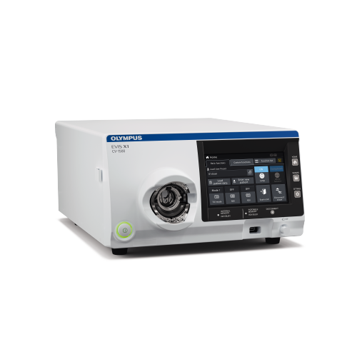
 CV-1500_Brochure_EN


 中文
中文 English