 EndoscopyConsumables
 English
 中文
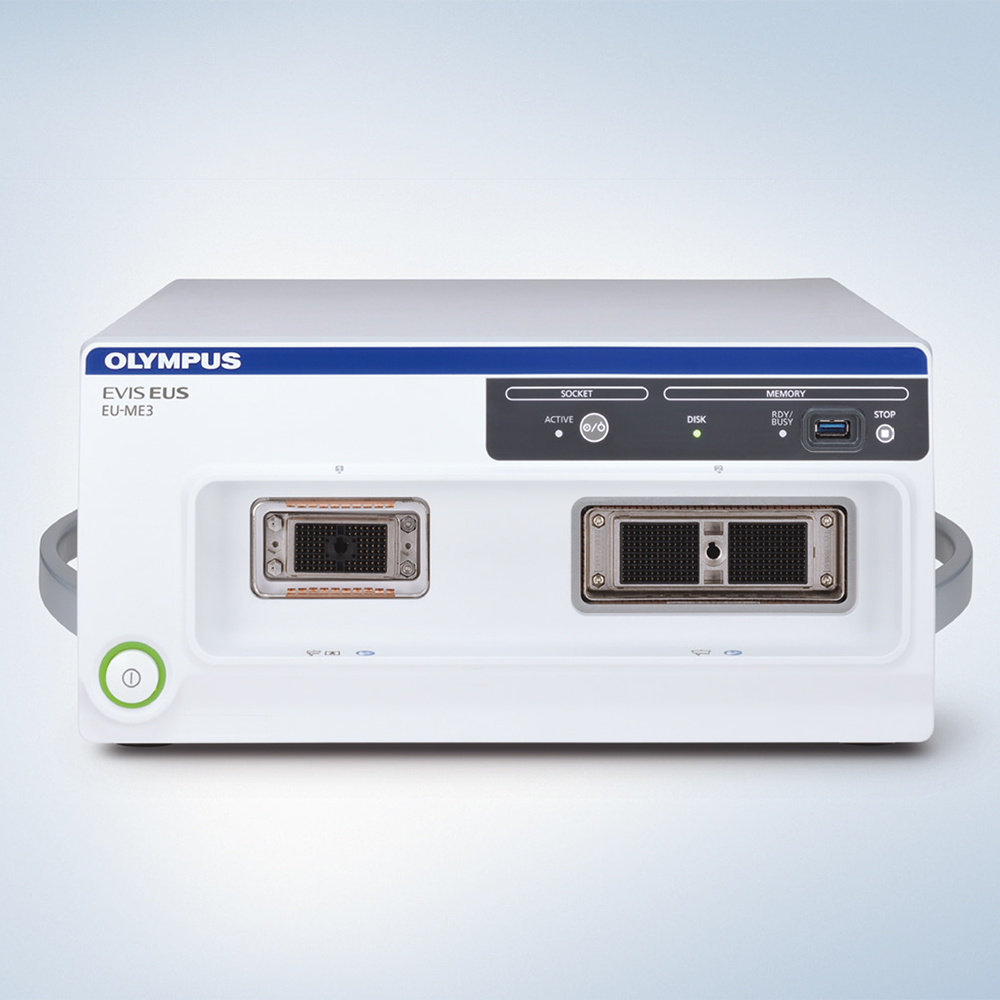
中文Olympus EU-ME3 Endoscopic Ultrasound Processor
The EU-ME3 is built to support more accurate diagnosis and treatment of hepatobiliary-pancreatic and lung diseases.
Phone:+86-21-54286005
Resources
 EU-ME3_Brochure_EN


 Microsystem
 Endoscopysystem
 Energysystem
 +86-21-54286005
 info@tenmed.net
 Room 602, Building 1, No. 111 Luxiang Road (Greenland Park Plaza), Baoshan District, Shanghai, China
The EU-ME3 is built to support more accurate diagnosis and treatment of hepatobiliary-pancreatic and lung diseases.
Phone:+86-21-54286005
EU-ME3_Brochure_EN
Advancing the Dimensions of Endosonography
With more functions, better visualization,and enhanced operability, the EU-ME3 expandsthe dimensions of endosonography.

Enhanced Visualization
Enhanced B-mode
The EU-ME3 provides outstanding image quality and functionality – compatible to a high-end ultrasound center – in a compact body. B-mode image quality has been substantially enhanced compared to our conventional processor (EU-ME2).

Improved Elastography
The EU-ME3 features an elastography function which visualizes the amount of strain in the tissue (tissue stiffness)during compression and retraction, making it possible to obtain more information about tissue properties.

Contrast Harmonic Echo (CHE)
Contrast Harmonic Echo (CHE) images harmonic components from ultrasound contrast agents.
The newly added C-THE mode images signals from biological tissue and the contrast.

Tissue Harmonic Echo (THE)
When ultrasound waves are propagated through tissue, distortion is produced and harmonic components are generated. The Tissue Harmonic Echo (THE) mode uses these components to build an image of the targeted area, providing a more detailed granular depiction. Advantages of harmonic imaging include improved resolution,improved signal-to-noise ratio, and fewer artifacts.
Doppler Modes
The EU-ME3 offers three basic Doppler modes to distinguish blood flow more clearly - Color Flow, Power Flow,and Pulsed Wave Doppler (PWD). Doppler modes can be used to support safer procedures, benefitting both the patient and the physician.
In addition to the three basic Doppler modes, the EU-ME3 also features H-Flow. H-Flow is a more sensitive Doppler mode that shows directional blood flow with less blooming. It is especially useful for imaging small vessels around the tip of the echoendoscope.


Color Flow Power Flow


Pulsed Wave Doppler H-Flow
Enhancing Functionality
Shear Wave Quantification (SWQ)
SWQ provides an absolute value of tissue stiffness within a region of interest. It performs this quantitative tissue assessment by calculating the propagation velocity of shear waves, generated from a push-pulse.

Elastography (i-ELST)
i-ELST is a new technology incorporated into the EU-ME3 that makes it easier to display elastic images, even when displacement due to pulsation is modest.
s-FOCUS
The EU-ME3 is equipped with an s-FOCUS mode that reduces the change in resolution with distance from the ultrasound transducer surface. s-FOCUS eliminates the need to manually adjust the focal zones during the procedure.

Keyboard Usability
The keyboard was designed with a simple layout in mind and includes a user-friendly built-in touch panel, LED backlit keys and a trackpad for ease of use and cleaning. The larger LCD touch panel allows for a greater range of functions to be displayed at one time.

Ease of Targeting
The position and size of the Doppler region of interest (ROI) can be conveniently adjusted with a trackpad or buttons on the touch panel.
Wide Range of Compatibility
Integrating both electronic and mechanical scanning technologies, the EU-ME3 is compatible with echoendoscopes and miniature probes, creating a total endosonography solution for a full range of applications.

Customizable Features
Software options are available to meet the needs of any facility. Because the functions are optional, you can select and add the necessary functions according to your needs and budget.

Comparison of Ultrasound Functions
| EU-ME2 |
EU-ME2 PREMIER |
EU-ME2 PREMIER PLUS |
EU-ME3 | |
| B-mode | ✓ | ✓ | ✓ | ✓ |
| THE (Tissue Harmonic Echo) | ✓ | ✓ | ✓ | |
| Flow | ✓ | ✓ | ✓ | ✓ |
| PWD (Pulsed Wave Doppler) | ✓ | ✓ | ✓ | ✓ |
| CHE (Contrast Harmonic Echo) | ✓ | ✓ |
✓ (Software Option) |
|
| Elastography | ✓ |
✓ (Software Option) |
||
| SWQ (Shear Wave Quantification)* |
✓ (Software Option) |
* For GI. Only compatible with GF-UCT180/260 and GF-UE190/290.
| Power Supply | Voltage |
220 – 240 V AC |
|
| Voltage fluctuation | Within ±10% | ||
| Frequency |
50/60 Hz |
||
| Frequency fluctuation | Within ±1 Hz | ||
| Consumption electric power |
340 VA |
||
| Size | Dimensions | Main unit | 371 (W) × 175 (H) × 480 (D) mm 445 (W) × 184 (H) × 530 (D) mm (max.) |
| Keyboard | 392 (W) × 39 (H) × 210 (D) mm | ||
| Weight | Main unit | 21.5 kg (without software option case) 21.8 kg (with software option case) |
|
| Keyboard |
2.5 kg |
||
| Classification | Type of protection against electric shock |
Class I | |
| Degree of protection against electric shock or applied part |
TYPE BF applied part where no classification mark appears, the device is a TYPE BF applied part. |
||
| Degree of protection against explosion |
The Ultrasound Center should be kept away from flammable gases. | ||
| Ultrasound Scanning Format | Mechanical scanning, electronic scanning | ||
| Mechanical Scanning | Display mode | B-mode | |
|
Scanning |
Radial scanning, helical scanning | ||
| Usable frequencies | 12 MHz, 20 MHz | ||
| Display range |
2,3,4,6,9,12 cm |
||
| Display processing |
Rotation |
Rotatable |
|
|
Display area |
Full circle, bottom sector, top sector, scroll | ||
|
Direction |
Normal/Inverse | ||
|
Cine memory |
Over 1,500 frames storable depending on the conditions. Cine review function |
||
|
3D |
3D display, MPR display | ||
|
Measurement |
Distance, area, circumstance | ||
| Electronic Scanning | Display mode | B-mode, FLOW mode, PW mode, CHE mode, ELST mode | |
|
Scanning |
Radial scanning, curved linear array scanning | ||
| Usable frequencies |
5 MHz,6 MHz,7.5 MHz,10 MHz,12 MHz |
||
| Display range |
2,3,4,5,6,7,8,9,12 cm |
||
| Display processing |
Rotation | Rotatable during radial scanning | |
| Display area | Radial: Full circle, bottom sector, top sector, scroll, Curved linear array: Fixed |
||
| Direction | Normal/Inverse | ||
|
Cine memory |
Over 2,000 frames storable depending on the conditions. Cine review function |
||
| Focus | Auto preset | s-FOCUS, AUTO, MANUAL | |
| Focus settings | Focus location and Focus number adjustable. | ||
| FLOW mode | COLOR-FLOW mode, POWER-FLOW mode, H-FLOW mode | ||
| PW mode | B+PW, COLOR+PW, POWER+PW, H-FLOW+PW | ||
|
Measurement |
Distance, area, circumstance, PW measurement | ||
| THE mode |
THE-P,THE-R |
||
| CHE mode (Software Options) |
Display pattern | CHE, C-THE | |
| Preset (CH agent type) |
2 types (Low acoustic pressure, Middle acoustic pressure), selectable | ||
| Frequency selection |
2 types (CHE-P, CHP-R) | ||
| ELST mode (Software Options) |
Pressurization guide |
Pressurization bar, Strain graph | |
| Strain ratio | Measures strain or ratio of strain of 2 areas. | ||
| SWQ (Software Options) | Calculates and displays transmission speed and elasticity of shear wave in ROI. |
||
| Recording Data |
Data format | Movie data |
Avi |
| Ancillary Equipment |
Keyboard | Built-in track pad and touch panel. | |
| Recording device | DVR | ||
| Video system center |
Monitor display selection |
Endoscopic/ultrasound image | |
| Sub screen | Endoscopic image can be displayed in sub screen. | ||
| Patient data | Patient data can be shared with video system center. | ||


 中文
中文 English